Table Of Content
This is the largest number of hair follicles you will ever have. As long as new hair cells continue to grow in the hair bulb, the hair continues to grow longer. At any point in time, about 90 percent of a person’s total amount of hair is in this growth phase. The first diagram of the hair shows the full structure of the skin.
Structure of hair shaft under a microscope
The first is the anagen phase, during which cells divide rapidly at the root of the hair, pushing the hair shaft up and out. The length of this phase is measured in years, typically from 2 to 7 years. The catagen phase lasts only 2 to 3 weeks, and marks a transition from the hair follicle’s active growth. Finally, during the telogen phase, the hair follicle is at rest and no new growth occurs. At the end of this phase, which lasts about 2 to 4 months, another anagen phase begins. The basal cells in the hair matrix then produce a new hair follicle, which pushes the old hair out as the growth cycle repeats itself.
Structure of hair follicle under a microscope
A club hair may fall out during catagen or telogen, or as it is pushed out by the new hair in the next anagen phase. The cylindrical shaft of the hair under a microscope shows three layers (medulla, cortex, and cuticle) of keratinized cells. Again, the terminal end of the hair follicle shows an expanded hair bulb composed of connective tissue papilla and hair root. Hair under a microscope shows a hair follicle and a cylindrical hair shaft. In a hair shaft, you will find columns of keratinized cells organized into three layers – medulla, cortex, and cuticle.
How can you tell if the hair is under a microscope?
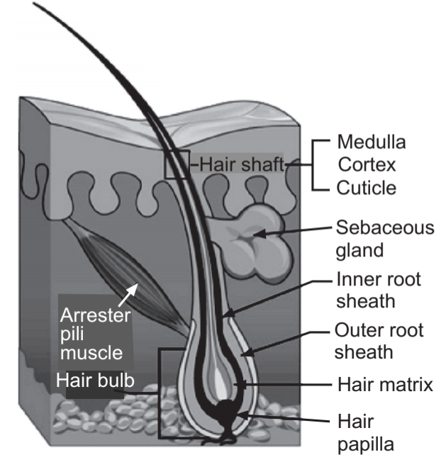
The most important one of these glands is the sebaceous gland, which produces and secretes the natural oils lubricating hairs, namely sebum. So, the hair under a light microscope shows two important features – hair shaft and hair follicle. The important features of the hair shaft are the cuticle, cortex, and medulla, which you might identify with the help of a light microscope.
The 8 Best Hair Clippers for Men 2024, Tested and Reviewed By Barbers - Men's Health
The 8 Best Hair Clippers for Men 2024, Tested and Reviewed By Barbers.
Posted: Fri, 08 Sep 2023 07:00:00 GMT [source]
Here, it shows the different epidermis layers and dermis of animal skin with a hair shaft and follicle. The contraction of the arrector pili muscle pressed upon the sebaceous gland helps them secret within the hair follicle. You will quickly identify and differentiate the animal hair from human hair under a light microscope with the help of their cuticle and medulla features. Generally, the animal hair contains a continuous or stacked medulla in its hair shaft. You will learn more about the microscopic features of the sebaceous glands of the hair follicle in the next section of this article. Sebaceous are the branched acinar holocrine glands that attach to the hair follicle.
You will also find more oval structures in the shaft of cattle or goat hair than in humans. It is very easy to visualize the scales and medulla of a hair shaft. Simply, you should follow a simple procedure and need some instruments. You will find the uniserial, multiserial ladder, vacuolated, and lattice medulla in different animal species.

The cellular or vacuolated medulla are most common in many animal species. All these uniserial, multiserial, cellular or vacuolated, and lattice medulla are shown in the hair labeled diagrams. All these types of medulla of hair are shown on the labeled diagrams. The appearance of the medulla of different types of hair is different.
10 Best Manscaping Tips - How to Manscape and Safely Shave Balls - Men's Health
10 Best Manscaping Tips - How to Manscape and Safely Shave Balls.
Posted: Thu, 20 Jul 2023 07:00:00 GMT [source]
Hair and Scalp Disorders
But the root of the cat hair is elongated and does not possess any distinct shape. Let’s see some of the common and important features of the hairs from other different parts of an animal’s body. The hair may be straight, curly, and kinly in different animal species.
Structure and Function
It has several types of stem cells, which develop into specialized cells and can renew themselves over a long period of time. The base of the root of a cat’s hair shows a frayed fibrils appearance. In the labeled diagram of the cat’s hair, I tried to show some of the important features from the hair shaft and follicle. You will find numerous blood vessels and numerous connective tissue fibers in the connective tissue of the hair follicle. These numerous vessels and fibers form the basket-like network around the lower end of the hair follicle. The outer layer of the hair follicle starts from the cuboidal cells of the outer layer and continues with the stratum spinosum of the skin.
Telogen effluvium describes a disruption in the normal hair growth and rest cycles that results in excessive shedding and hair loss. This condition can be precipitated by physical stress, such as surgery or malnutrition, or by medical exposure, such as chemotherapy and common medications. The hair shaft is the portion of your hair that projects from your scalp. The root of each hair is located in a follicle, which is embedded in the skin and nourished by the blood vessels in the dermis (see diagram). In a young adult, scalp follicles typically spend 6 to 8 years in anagen, 2 to 3 weeks in catagen, and 1 to 3 months in telogen. Scalp hairs grow at a rate of about 1 mm per 3 days (10–18 cm/yr) in the anagen phase.
You will see a glassy membrane in the diagram that separates the inner root layer from the outer root layer. The hair bulb represents the hair matrix, and hair follicles stem cells. These hair matrices and stem cells are responsible for forming the hair. The stem cells proliferate, move upwards, and gradually become keratinized to produce the hair. The outer root layer of the hair follicle is continuous with the epidermis. You will find a glassy basement membrane that separates this outer root layer from the surrounding connective tissue.

FGF5 is a key inducer of catagen and FGF5-deficient mice have a prolonged anagen phase. In addition to FGF5, TGF-β1, IL-1b, the neurotrophins NT-3, NT-4 and BMP2/4 and TNF-α have been described to induce catagen [36]. Moreover the follicular papilla is an essential source of growth factors [1, 3, 16, 28]. Through the anagen I–V, hair stem cells proliferate, encloses the dermal papilla, grow downwards to the skin and begin to proliferate hair shaft and IRS, respectively. This phase can last up to 6–8 years in hair follicles [1, 11, 18].
During catagen the proximal of the hair shaft is keratinized and forms the club hair, whereas the distal part of the follicle is involuted by apoptosis [16, 38]. In humans, it pulls the follicles into a vertical position and causes “goose bumps,” but serves no useful purpose. The hair follicles in your skin contain living cells to allow your hair to grow. The shaft—the part of the hair we see—is made up of dead cells and consists of three different layers. As a piece of hair grows, it goes through three phases before it sheds and a new one grows. Again, this diagram shows different layers of rounded nucleated cells that form the outer root layer of the hair follicle.
But, these spinous or petal-like scales are not found in the human hair shaft. The hair shaft under a microscope shows three different distinct layers – an inner medulla, a cortex, and an outer cuticle. The structure of a pencil may be a good analogy for the structure of a hair shaft. The cells rupture to secrete an oily secretion into the lumen of the hair follicle. Again, the ruptured cells are continuously replaced by stem cells located at the edges of the glands. Again, let’s see the cross-section of the hair follicles and try to identify the following features from the labeled diagram.
From bleach blonde hair color to the trendiest hair accessories we will try anything to make our crowns stand out. Our hair grows in so many different shades, places, hair textures, and types, that everyone’s mane is naturally unique. But when it comes to hair anatomy, all of our strands are essentially the same. A piece of hair may look simple, but it’s one of the body's most complicated structures. The hair follicle is the part below the skin, and the hair shaft is what you see above your skin.
In hair follicles, 5-alpha reductase is found in the sebaceous glands, dermal papilla, inner root sheath and outer root sheath (2). And androgen receptor sites are found in dermal papilla cells and sebaceous glands (3). Again, the hair matrix stem cells help form the inner and outer root layer of the hair follicle. There is a dermal papilla at the base of the hair bulb and remains in the skin’s dermis. Hair texture (straight, curly) is determined by the shape and structure of the cortex, and to the extent that it is present, the medulla. The shape and structure of these layers are, in turn, determined by the shape of the hair follicle.


No comments:
Post a Comment